Negative Stain Transmission Electron Microscopy
This method is routinely used in screening experiments to assess sample quality. It is also used to answer specific questions concerning, e.g., protein-protein interactions and protein assembly; antibodies can be visualized when bound to their antigen.
The sample is embedded in a layer of a heavy metal salt. This scatters a large number of electrons and looks dark on the images.
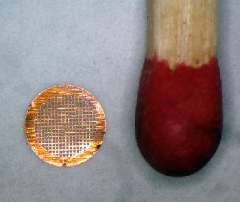
Copper EM-grid, 3mm in diameter compared to the head of a match stick.
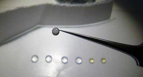
Negative staining: The sample-loaded grid is washed on a droplet of water (colorless) and stained on droplets of a heavy metal salt, here 2% uranyl acetate (yellow).
The magnification series is continued in the slider below. The thin carbon spanning a hole in the perforated layer and the negatively stained protein complexes on it, become increasingly visible.
Images courtesy of Philippe Ringler, University of Basel.